View Abnormal Bladder Ultrasound Images Images. Philips lumify handheld ultrasound is available for medical professionals to use in many different settings. Bladder ultrasound education showing how to, scanning protocol, normal anatomy, anatomic variants, volume, residual, ureteric jet, haematuria.
Pocus Gallery Renal Fellow Network from www.renalfellow.org The procedure allows your doctor to view images of your gallbladder to inform their diagnosis. The bladder wall thickness was measured from a zoomed image of the transverse plane of the voided bladder at 3 points: Ultrasound imaging may be done in the health provider's office, in the hospital, or in an outpatient facility.
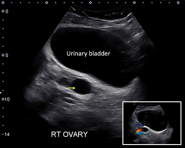
This is especially important when a patient is not sexually active or a virgin, because the most pelvic ultrasounds are two part exams:
The ureterocele is seen partially distended and also seen in the collapsing stage as the pressure builds up within the sac (of. Bladder ultrasounds can be used independently for volume measurements or in conjunction with other exams such as fast, renal studies, and pelvic ultrasounds. It is used to help diagnose pain or distention for ultrasound of the kidneys, you may be asked to drink four to six glasses of liquid about an hour before the test to fill your bladder. The procedure allows your doctor to view images of your gallbladder to inform their diagnosis.